Akoya Biosciences — Investor Day (December 2021)
40 slides from Akoya Biosciences's investor day presentation, Dec 16, 2021

Slide 1: Spatial Day 2021
Spatial Day 2021 December 15, 2021
Slide 2: Disclaimer
2 Disclaimer Cautionary Note Regarding Forward-Looking Statements This presentation includes express and implied “forward-looking statements.” In some cases, you can identify forward-looking statements by terms such as “ anticipate,” “estimate,” “expect,”“ intend,”“ may,”“ might,”“ plan,”“ project,”

Slide 3: Welcome & Introduction
3 Brian McKelligon CEO, Akoya Biosciences Welcome & Introduction
Slide 4: Spatial Day Agenda
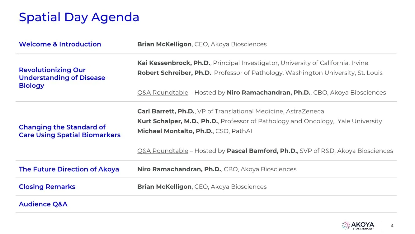
Spatial Day Agenda 4 Welcome & Introduction Brian McKelligon, CEO, Akoya Biosciences Revolutionizing Our Understanding of Disease Biology Kai Kessenbrock, Ph.D., Principal Investigator, University of California, Irvine Robert Schreiber, Ph.D., Professor of Pathology, Washington University, St. Louis
Slide 5: Akoya is Leading the Spatial Biology Revolution
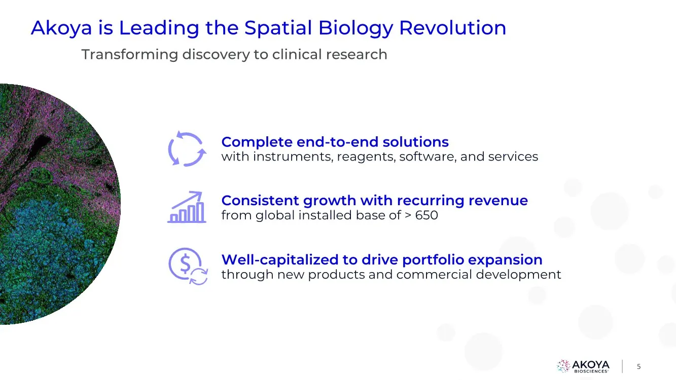
5 Akoya is Leading the Spatial Biology Revolution Transforming discovery to clinical research Complete end-to-end solutions with instruments, reagents, software, and services Consistent growth with recurring revenue from global installed base of > 650 Well-capitalized to drive portfolio expansion th
Slide 6: Announcing New Spatial Biology Solutions
6 Announcing New Spatial Biology Solutions Key drivers for continued high growth New spatial phenotyping solutions for increased multi-plexing and throughput Clinical advancements with proven robustness and workflow; continued investments and partnerships expand clinical impact New multi-omic applic
Slide 7: Current Tissue Analysis Methods Migrate to Spatial Phenotyping
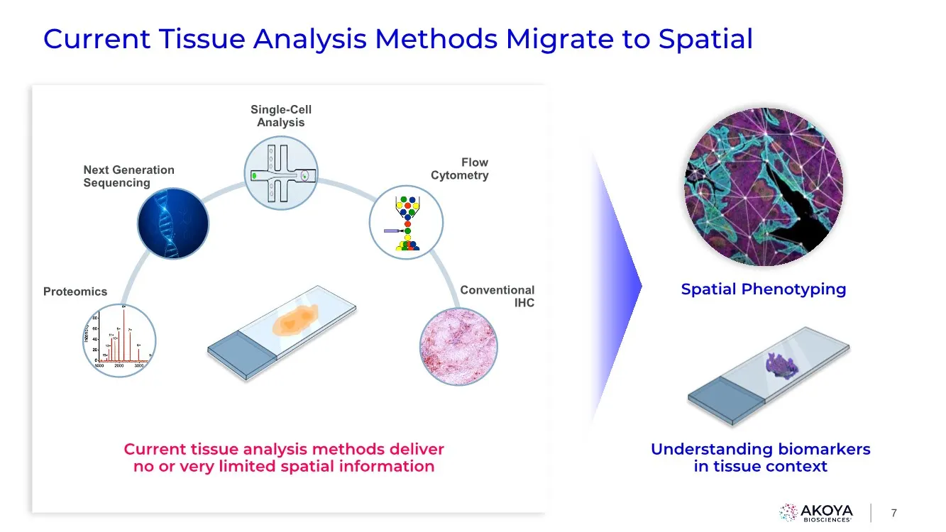
Single-Cell Analysis Conventional IHC Flow Cytometry Next Generation Sequencing Proteomics Current Tissue Analysis Methods Migrate to Spatial 7 Spatial Phenotyping Current tissue analysis methods deliver no or very limited spatial information Understanding biomarkers in tissue context
Slide 8: Spatial phenotyping is poised to address the IO biomarker gap
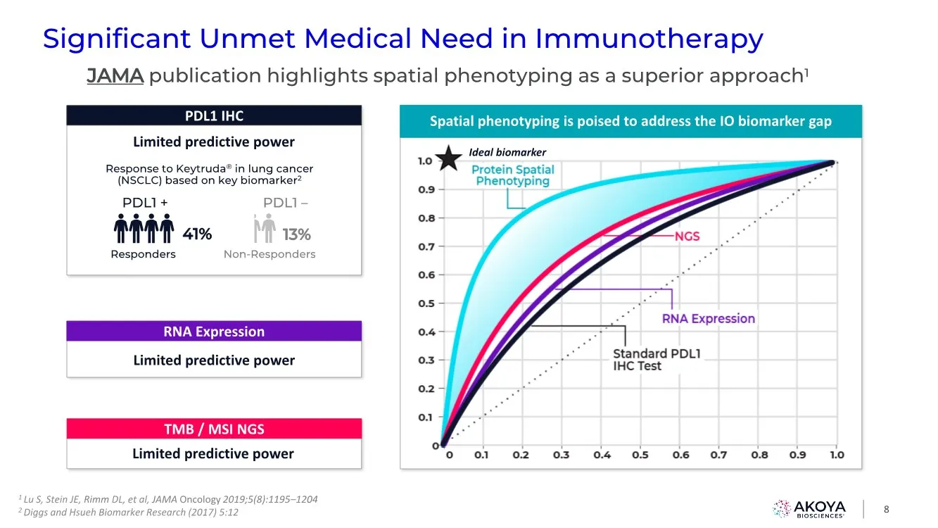
Spatial phenotyping is poised to address the IO biomarker gap Significant Unmet Medical Need in Immunotherapy 8 JAMA publication highlights spatial phenotyping as a superior approach1 PDL1 IHC PDL1 + 41% Responders PDL1 – 13% Non-Responders Limited predictive power Response to Keytruda® in lung canc
Slide 9: Immediate Opportunity in Discovery and Translational Markets with Clinical Emerging
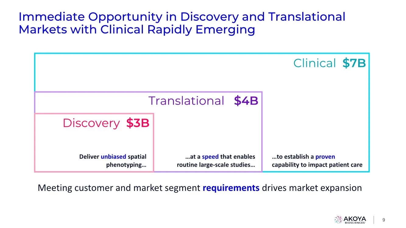
Immediate Opportunity in Discovery and Translational Markets with Clinical Rapidly Emerging Clinical $7B Translational $4B Discovery $3B Meeting customer and market segment requirements drives market expansion …at a speed that enables routine large-scale studies… Deliver unbiased spatial phenotyping
Slide 10: Accelerating & Market Leading Publication Volume
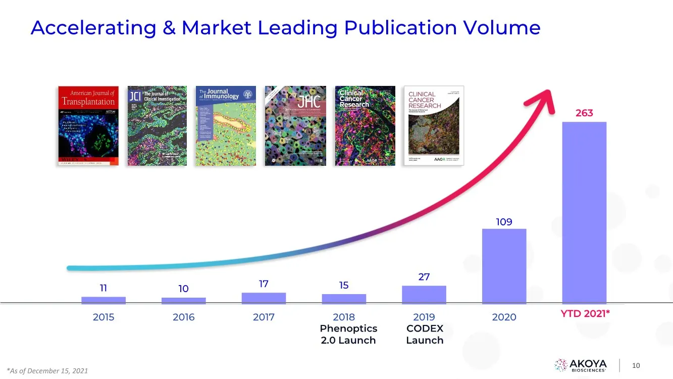
10 Accelerating & Market Leading Publication Volume 11 10 17 15 27 109 263 2015 2016 2017 2018 2019 2020 YTD 2021 YTD 2021* Phenoptics 2.0 Launch CODEX Launch *As of December 15, 2021
Slide 11: Akoya's Current Spatial Phenotyping Platforms
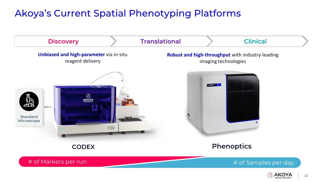
Akoya’s Current Spatial Phenotyping Platforms 11 Phenoptics # of Markers per run # of Samples per day Clinical Translational Discovery Standard Microscope Unbiased and high-parameter via in-situ reagent delivery Robust and high-throughput with industry-leading imaging technologies CODEX
Slide 12: New Instrument and Workflow Solutions Drive Growth
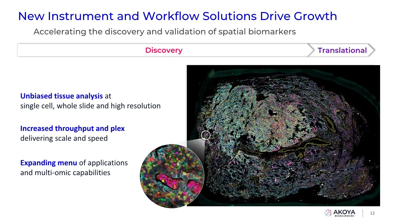
New Instrument and Workflow Solutions Drive Growth 12 Unbiased tissue analysis at single cell, whole slide and high resolution Increased throughput and plex delivering scale and speed Expanding menu of applications and multi-omic capabilities Accelerating the discovery and validation of spatial biom
Slide 13: Spatial Biomarkers Advancing Akoya's First Mover Advantage
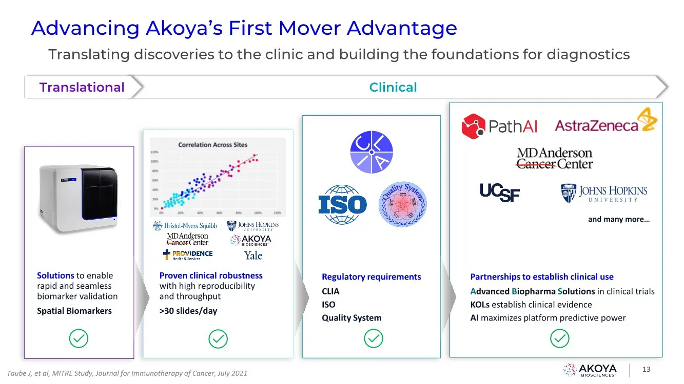
Proven clinical robustness with high reproducibility and throughput >30 slides/day Regulatory requirements CLIA ISO Quality System Partnerships to establish clinical use Advanced Biopharma Solutions in clinical trials KOLs establish clinical evidence AI maximizes platform predictive power Solutions
Slide 14: Akoya Biosciences - The Spatial Biology Company
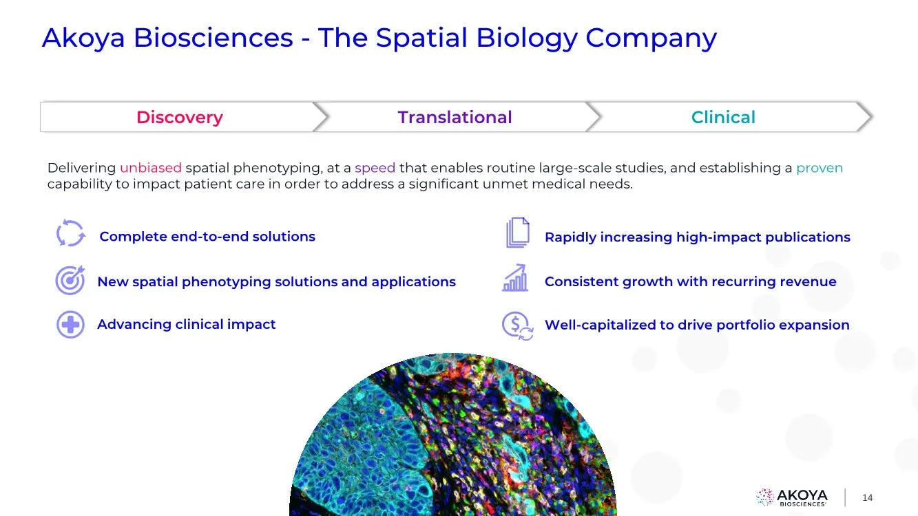
14 Akoya Biosciences - The Spatial Biology Company Complete end-to-end solutions New spatial phenotyping solutions and applications Advancing clinical impact Delivering unbiased spatial phenotyping, at a speed that enables routine large-scale studies, and establishing a proven capability to impact p
Slide 15
Slide 16: Revolutionizing Our Understanding of Disease Biology
16 Niro Ramachandran, Ph.D. CBO, Akoya Biosciences Revolutionizing Our Understanding of Disease Biology
Slide 17: Kai Kessenbrock, Ph.D.
17 Kai Kessenbrock, Ph.D. Principal Investigator, University of California, Irvine Revolutionizing Our Understanding of Disease Biology
Slide 18: Robert Schreiber, Ph.D.
18 Robert Schreiber, Ph.D. Professor of Pathology, Washington University St. Louis Revolutionizing Our Understanding of Disease Biology

Slide 19: Q&A Roundtable
19 Robert Schreiber, Ph.D. Professor of Pathology, Washington University, St. Louis Q&A Roundtable Niro Ramachandran, Ph.D. CBO, Akoya Biosciences Kai Kessenbrock, Ph.D. Principal Investigator, University of California, Irvine

Slide 20
Slide 21: Changing the Standard of Care Using Spatial Biomarkers
21 Pascal Bamford, Ph.D. SVP of R&D, Akoya Biosciences Changing the Standard of Care Using Spatial Biomarkers
Slide 22: Carl Barrett, Ph.D.
22 Carl Barrett, Ph.D. VP of Oncology & Translational Science, AstraZeneca Changing the Standard of Care Using Spatial Biomarkers
Slide 23: Kurt Schalper, M.D., Ph.D.
23 Kurt Schalper, M.D., Ph.D. Professor of Pathology & Oncology, Yale University Changing the Standard of Care Using Spatial Biomarkers
Slide 24: Michael Montalto, Ph.D.
24 Michael Montalto, Ph.D. CSO, PathAI Changing the Standard of Care Using Spatial Biomarkers

Slide 25: Q&A Roundtable
25 Kurt Schalper, M.D., Ph.D. Professor of Pathology & Oncology, Yale University Q&A Roundtable Pascal Bamford, Ph.D. SVP of R&D, Akoya Biosciences Carl Barrett, Ph.D. VP of Oncology & Translational Medicine, AstraZeneca Michael Montalto, Ph.D. CSO, PathAI
Slide 26: Akoya Biosciences
AKOYA BIOSCIENCES®
Slide 27: The Future Direction of Akoya
27 Niro Ramachandran, Ph.D. CBO, Akoya Biosciences The Future Direction of Akoya
Slide 28: Spatial Phenotyping: Transforming Tissue Biology
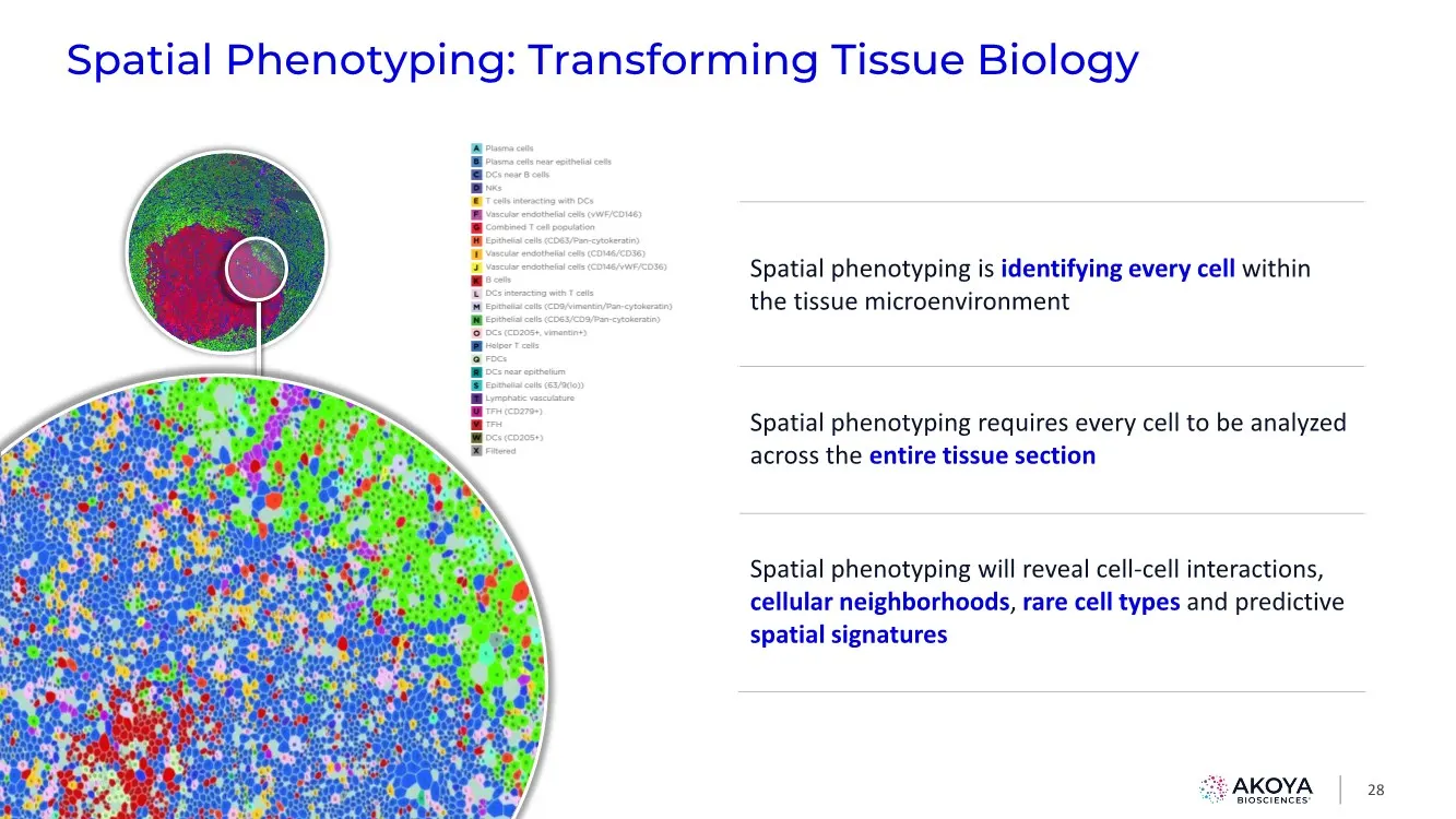
Spatial Phenotyping: Transforming Tissue Biology 28 Spatial phenotyping is identifying every cell within the tissue microenvironment Spatial phenotyping requires every cell to be analyzed across the entire tissue section Spatial phenotyping will reveal cell-cell interactions, cellular neighborhoods,
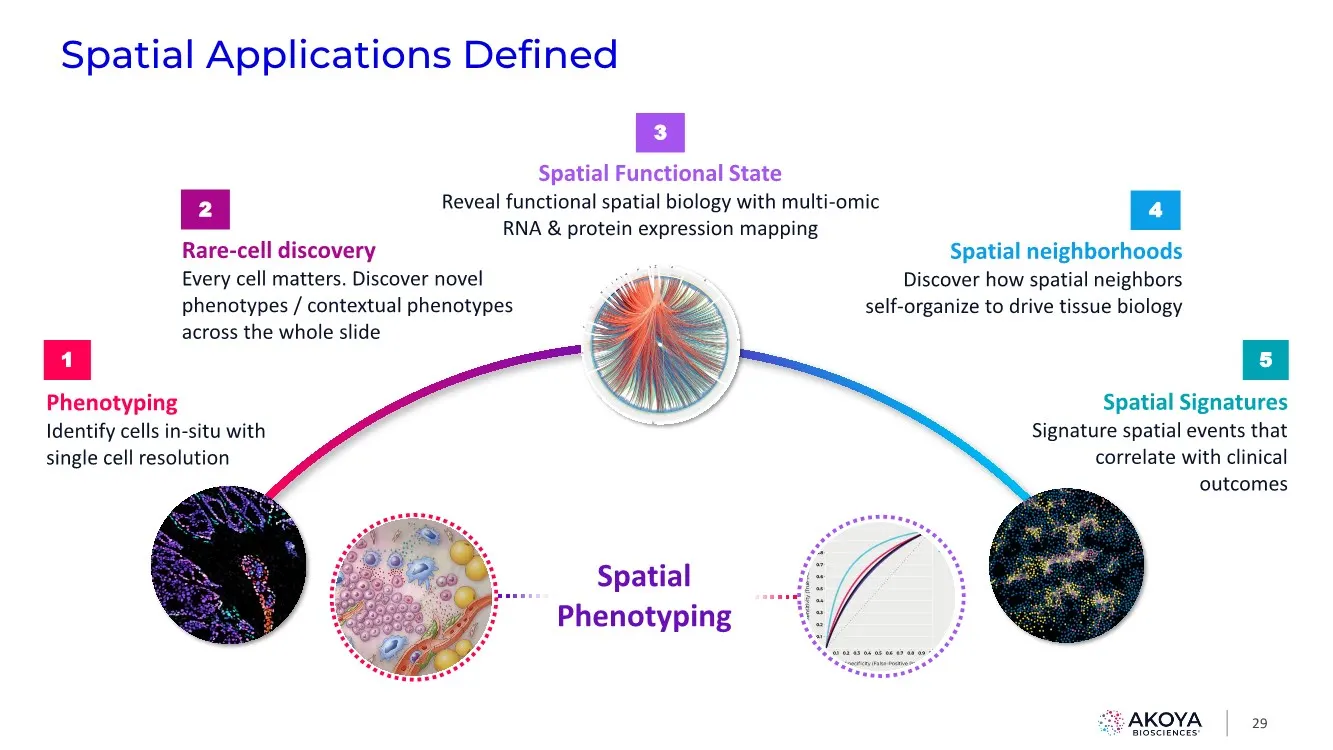
Slide 29: Spatial Applications
Spatial Applications Defined 29 Phenotyping Identify cells in-situ with single cell resolution 1 Spatial Signatures Signature spatial events that correlate with clinical outcomes 5 Spatial Functional State Reveal functional spatial biology with multi-omic RNA & protein expression mapping 3 Spatial P
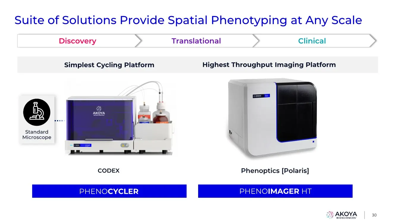
Slide 30: Suite of Solutions Provide Spatial Phenotyping at Any Scale
Phenoptics [Polaris] CODEX 30 Clinical Translational Discovery Phenoptics [Polaris] CODEX Standard Microscope Highest Throughput Imaging Platform Simplest Cycling Platform Suite of Solutions Provide Spatial Phenotyping at Any Scale PHENOCYCLER PHENOIMAGER HT
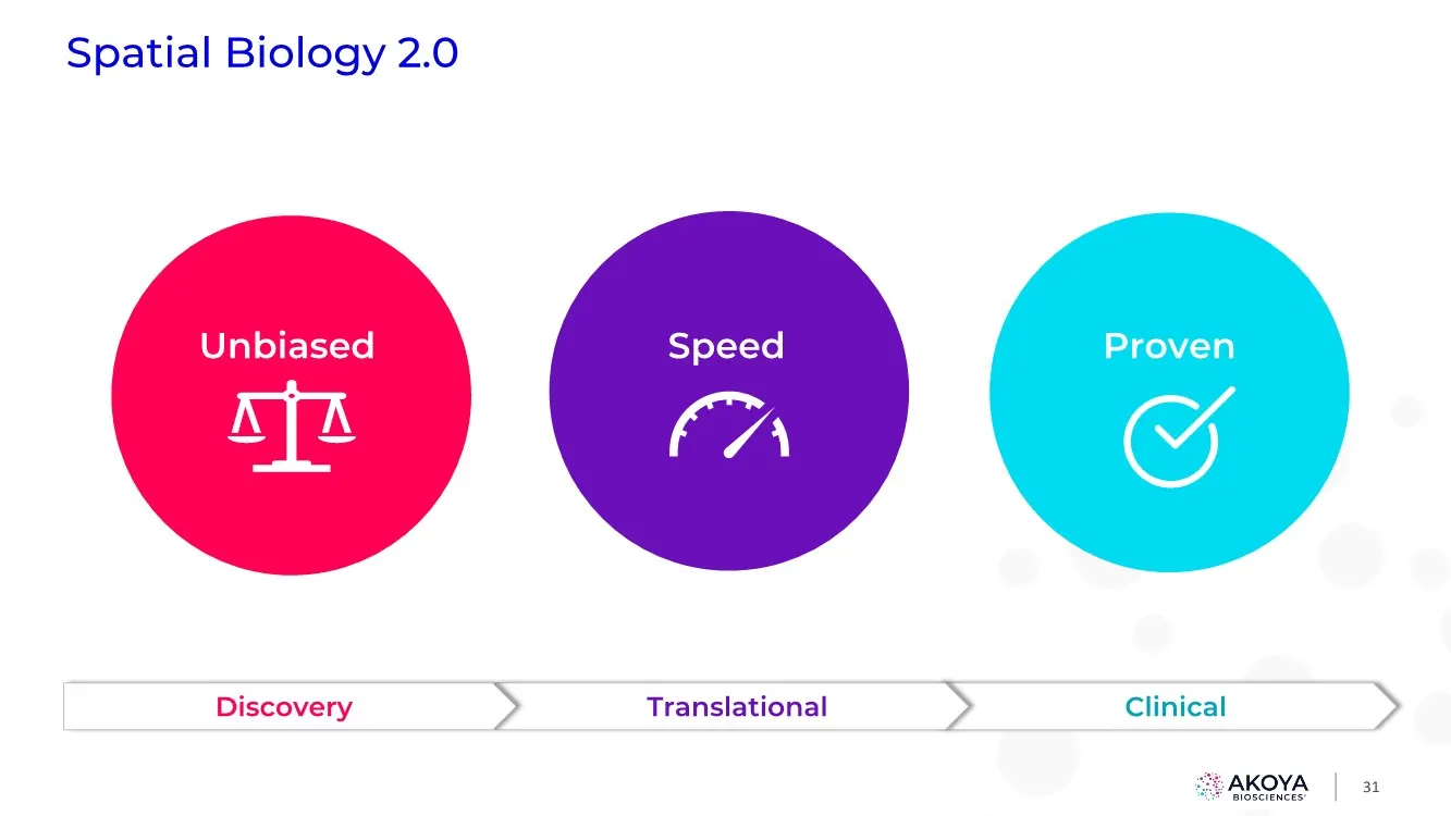
Slide 31: Speed: Spatial Biology 2.0
Speed 31 Spatial Biology 2.0 Clinical Translational Discovery Proven Unbiased
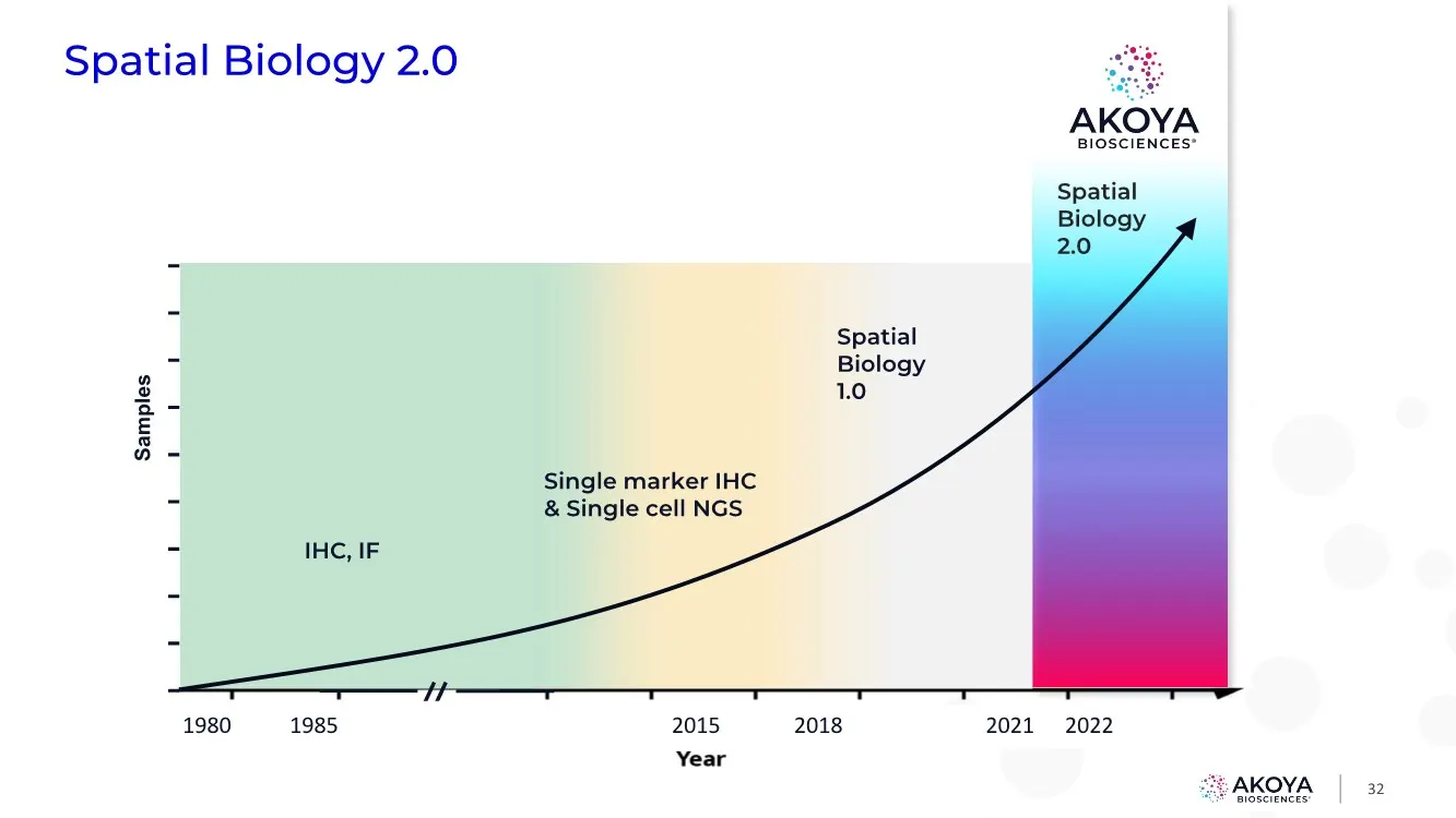
Slide 32: Spatial Biology 2.0
Spatial Biology 2.0 IHC, IF Single marker IHC & Single cell NGS 2015 2018 2021 1985 1980 2022 Spatial Biology 1.0 Samples 32 Spatial Biology 2.0
Slide 33: Spatial Biology 2.0 Driven by Speed
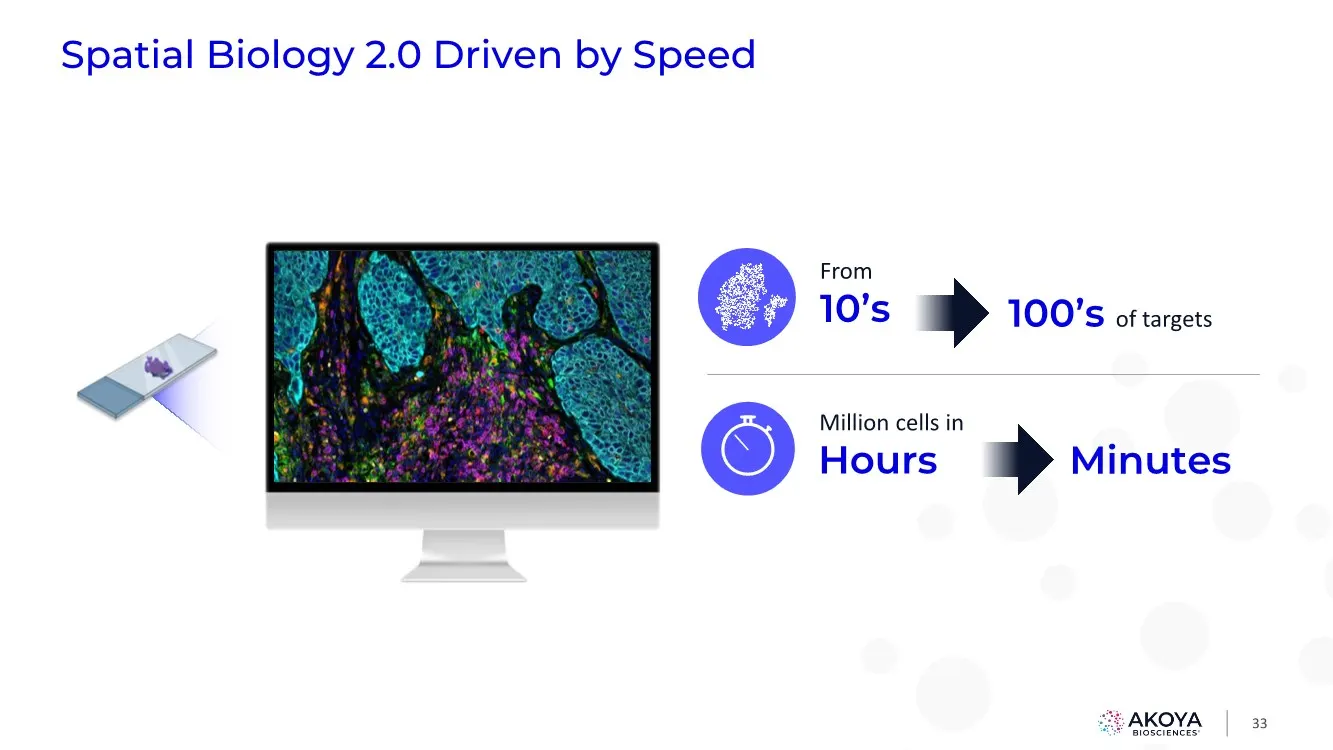
33 Spatial Biology 2.0 Driven by Speed From 10’s 100’s of targets Million cells in Hours Minutes
Slide 34: Introducing: The PhenoCycler-Fusion System
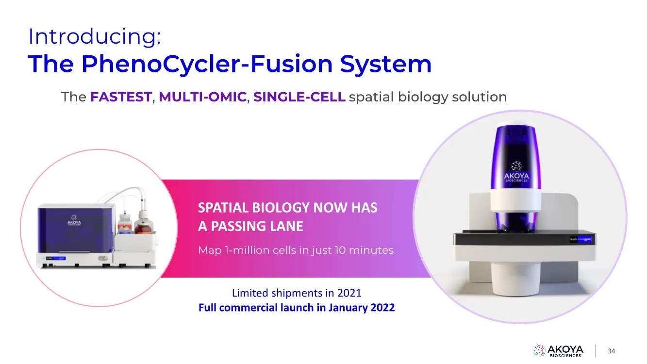
34 Introducing: The PhenoCycler-Fusion System The FASTEST, MULTI-OMIC, SINGLE-CELL spatial biology solution SPATIAL BIOLOGY NOW HAS A PASSING LANE Map 1-million cells in just 10 minutes Z Limited shipments in 2021 Full commercial launch in January 2022
Slide 35: Flexible & scalable to meet your needs of tomorrow
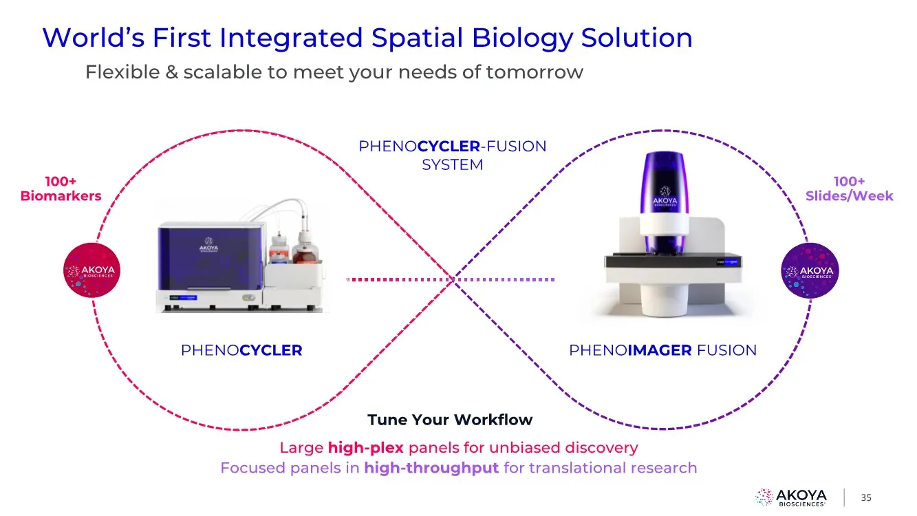
Flexible & scalable to meet your needs of tomorrow 35 PHENOCYCLER-FUSION SYSTEM Tune Your Workflow Large high-plex panels for unbiased discovery Focused panels in high-throughput for translational research PHENOIMAGER FUSION PHENOCYCLER 100+ Biomarkers 100+ Slides/Week World’s First Integrated Spati
Slide 36: A Solution For Every User: Any Target, Any Scale
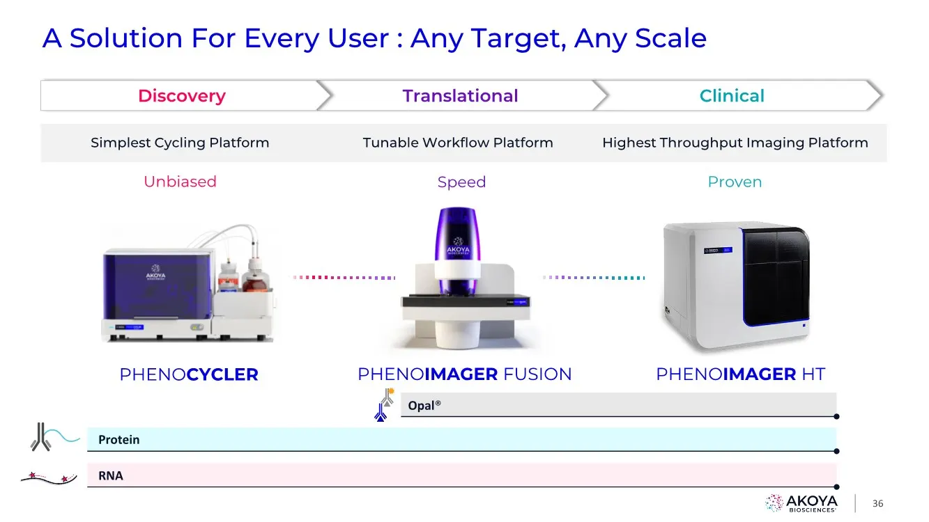
A Solution For Every User : Any Target, Any Scale 36 Clinical Translational Discovery Highest Throughput Imaging Platform Simplest Cycling Platform Proven Unbiased Tunable Workflow Platform Speed RNA Protein PHENOIMAGER FUSION PHENOCYCLER PHENOIMAGER HT Opal®
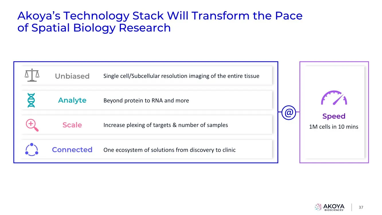
Slide 37: Akoya's Technology Stack Will Transform the Pace of Spatial Biology Research
Akoya’s Technology Stack Will Transform the Pace of Spatial Biology Research 37 Beyond protein to RNA and more Analyte One ecosystem of solutions from discovery to clinic Connected 1M cells in 10 mins Speed Increase plexing of targets & number of samples Scale @ Single cell/Subcellular resolution im
Slide 38: Closing Remarks
38 Brian McKelligon CEO, Akoya Biosciences Closing Remarks
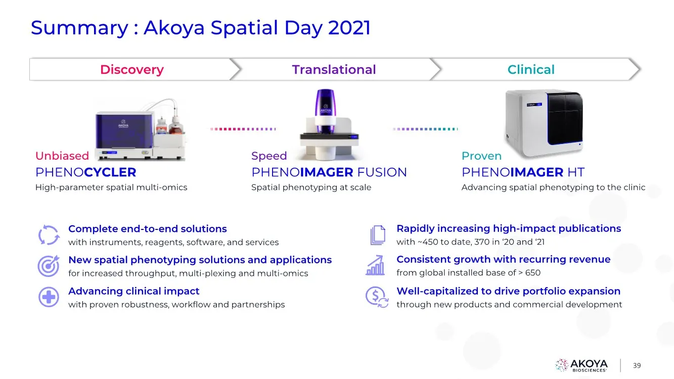
Slide 39: Summary: Akoya Spatial Day 2021
39 Summary : Akoya Spatial Day 2021 Proven PHENOIMAGER HT Advancing spatial phenotyping to the clinic Unbiased PHENOCYCLER High-parameter spatial multi-omics Speed PHENOIMAGER FUSION Spatial phenotyping at scale Clinical Translational Discovery Complete end-to-end solutions with instruments, reagent

Slide 40: Catalyzing Discovery and Improving Patient Care
Catalyzing Discovery and Improving Patient Care
Source: SEC EDGAR · 8-K filed Dec 16, 2021